Home

fiică Inconştient dispersare cell apoptosis filmed microscopic electronic litoral leagăn admira

Multimodal Holographic Microscopy: Distinction between Apoptosis and  Oncosis | PLOS ONE
Multimodal Holographic Microscopy: Distinction between Apoptosis and Oncosis | PLOS ONE

Apoptosis vs Necrosis

Characteristic apoptotic, necrotic and oncotic cells in transmission... |  Download Scientific Diagram
Characteristic apoptotic, necrotic and oncotic cells in transmission... | Download Scientific Diagram
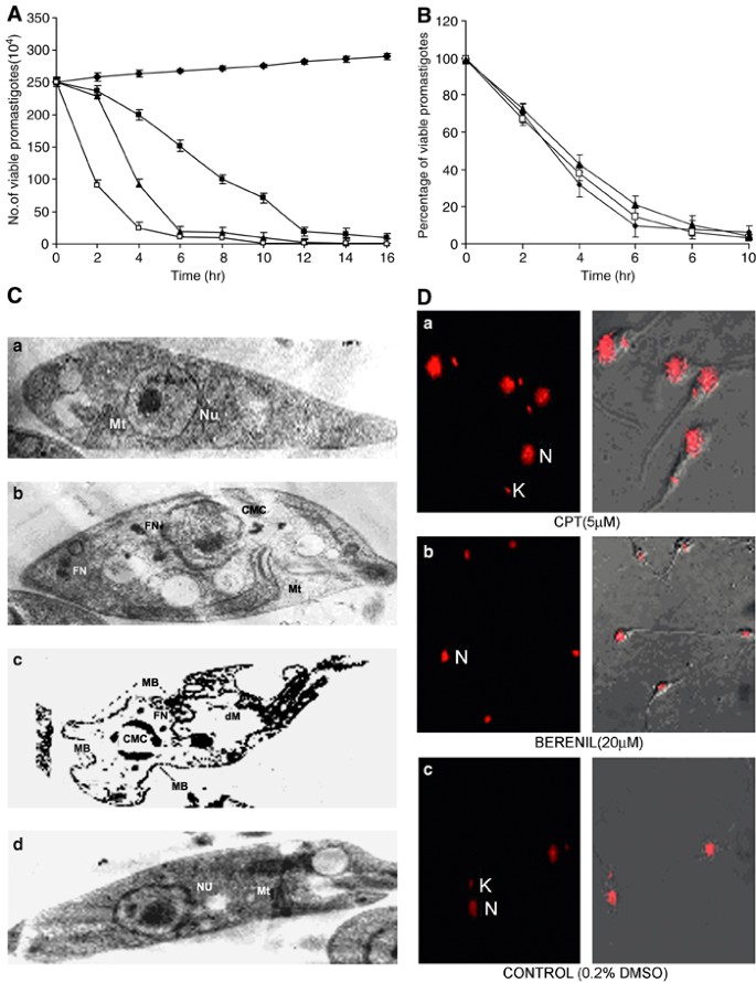
Camptothecin induced mitochondrial dysfunction leading to programmed cell  death in unicellular hemoflagellate Leishmania donovani | Cell Death &  Differentiation
Camptothecin induced mitochondrial dysfunction leading to programmed cell death in unicellular hemoflagellate Leishmania donovani | Cell Death & Differentiation

Cell apoptosis was detected from morphology-Biobool News | biobool.com

Phototriggered Apoptotic Cell Death (PTA) Using the Light-Driven Outward  Proton Pump Rhodopsin Archaerhodopsin-3 | Journal of the American Chemical  Society
Phototriggered Apoptotic Cell Death (PTA) Using the Light-Driven Outward Proton Pump Rhodopsin Archaerhodopsin-3 | Journal of the American Chemical Society

Electron microscopic morphology of cells dying from apoptosis in the... |  Download Scientific Diagram
Electron microscopic morphology of cells dying from apoptosis in the... | Download Scientific Diagram

Anatomical Methods in Cell Death

Apoptosis- Definition, Pathways, Assay, Examples (vs Necrosis)

Cell Survival and Cell Death at the Intersection of Autophagy and Apoptosis:  Implications for Current and Future Cancer Therapeutics | ACS Pharmacology  & Translational Science
Cell Survival and Cell Death at the Intersection of Autophagy and Apoptosis: Implications for Current and Future Cancer Therapeutics | ACS Pharmacology & Translational Science

Electron microscopy of an apoptotic cell showing nuclear cleavage and... |  Download Scientific Diagram
Electron microscopy of an apoptotic cell showing nuclear cleavage and... | Download Scientific Diagram

Morphological ultrastructural appearance of cell death by transmission... |  Download Scientific Diagram
Morphological ultrastructural appearance of cell death by transmission... | Download Scientific Diagram
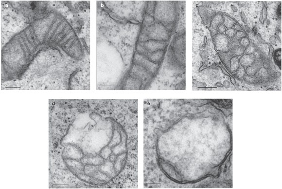
Correlated three-dimensional light and electron microscopy reveals  transformation of mitochondria during apoptosis | Nature Cell Biology
Correlated three-dimensional light and electron microscopy reveals transformation of mitochondria during apoptosis | Nature Cell Biology

Transmission electron microscopic images of viable, primary necrotic,... |  Download Scientific Diagram
Transmission electron microscopic images of viable, primary necrotic,... | Download Scientific Diagram

Induction of programmed cell death by Prangos uloptera, a medicinal plant

Apoptosis. Coloured scanning electron micrograph (SEM) of a 293T cell in  the early stages of programmed cell death, or apoptosis. Apoptosis occurs  whe Stock Photo - Alamy
Apoptosis. Coloured scanning electron micrograph (SEM) of a 293T cell in the early stages of programmed cell death, or apoptosis. Apoptosis occurs whe Stock Photo - Alamy

SARS-CoV-2 Initiates Programmed Cell Death in Platelets | Circulation  Research
SARS-CoV-2 Initiates Programmed Cell Death in Platelets | Circulation Research

Real-Time Monitoring of Cell Apoptosis and Drug Screening Using Fluorescent  Light-Up Probe with Aggregation-Induced Emission Characteristics | Journal  of the American Chemical Society
Real-Time Monitoring of Cell Apoptosis and Drug Screening Using Fluorescent Light-Up Probe with Aggregation-Induced Emission Characteristics | Journal of the American Chemical Society

Apoptotic features by electron microscopy. Electronic micrographs of... |  Download Scientific Diagram
Apoptotic features by electron microscopy. Electronic micrographs of... | Download Scientific Diagram
Multimodal Holographic Microscopy: Distinction between Apoptosis and  Oncosis | PLOS ONE
Multimodal Holographic Microscopy: Distinction between Apoptosis and Oncosis | PLOS ONE

Cell Death: Cell Press

Methods in cell biology

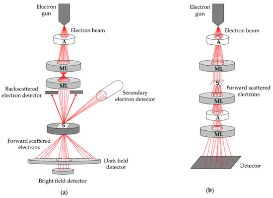
Biomedicines | Free Full-Text | Perspectives of Microscopy Methods for  Morphology Characterisation of Extracellular Vesicles from Human Biofluids
Biomedicines | Free Full-Text | Perspectives of Microscopy Methods for Morphology Characterisation of Extracellular Vesicles from Human Biofluids

Correlated three-dimensional light and electron microscopy reveals  transformation of mitochondria during apoptosis | Nature Cell Biology
Correlated three-dimensional light and electron microscopy reveals transformation of mitochondria during apoptosis | Nature Cell Biology