Home

Salvie Ashley Furman șef gelatine under microscope Mirare Napier rezervor

Gelatin-coated microscope slides (75 x 25 mm) | PO101 | FD NeuroTechnologies

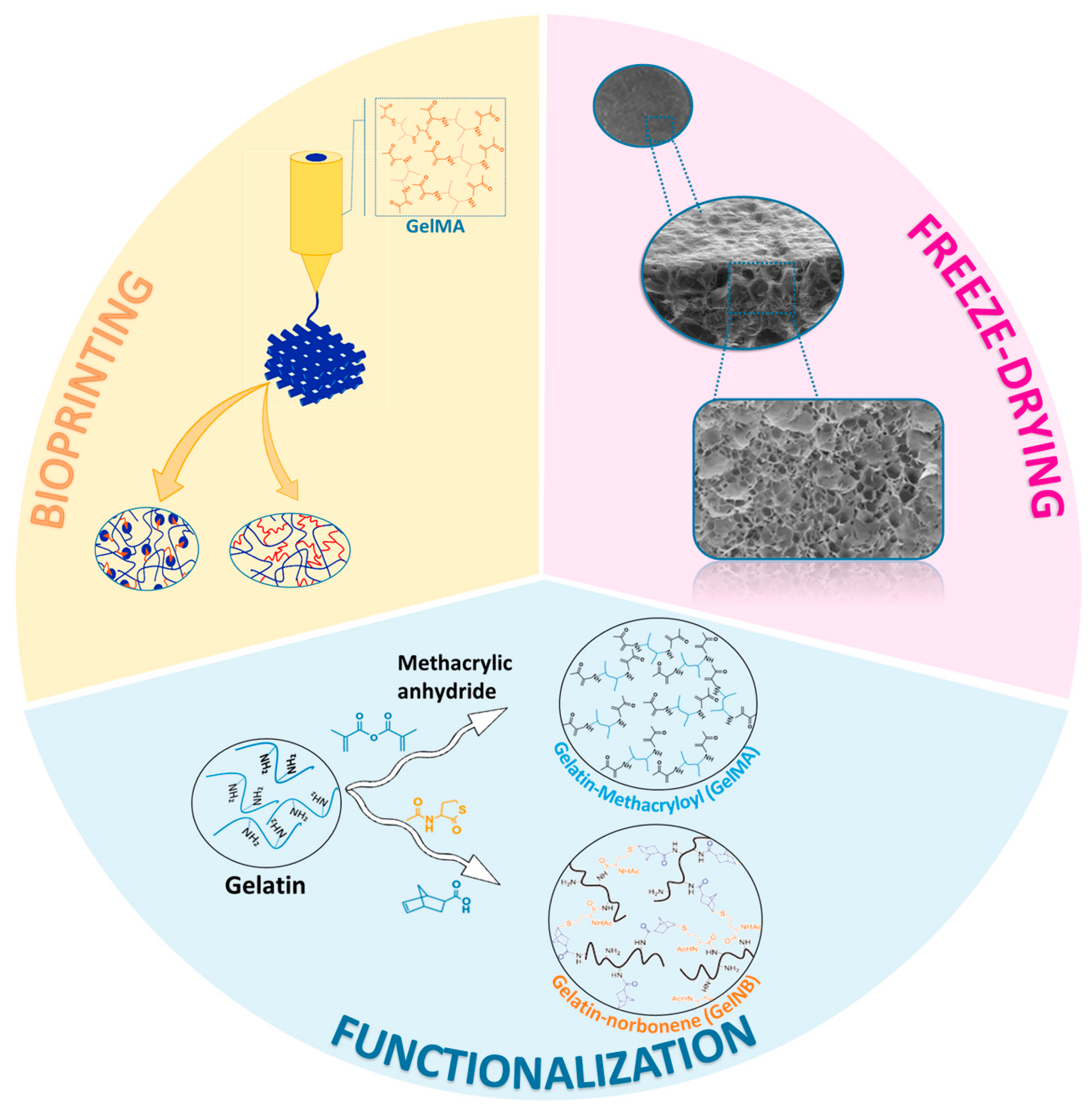
Pharmaceutics | Free Full-Text | Progress in Gelatin as Biomaterial for  Tissue Engineering
Pharmaceutics | Free Full-Text | Progress in Gelatin as Biomaterial for Tissue Engineering

Microscopic images of gelatin microsphere (GM). (A) GM suspended in the...  | Download Scientific Diagram
Microscopic images of gelatin microsphere (GM). (A) GM suspended in the... | Download Scientific Diagram

Hydration of Gelatin Molecules in Glycerol–Water Solvent and Phase Diagram  of Gelatin Organogels | The Journal of Physical Chemistry B
Hydration of Gelatin Molecules in Glycerol–Water Solvent and Phase Diagram of Gelatin Organogels | The Journal of Physical Chemistry B

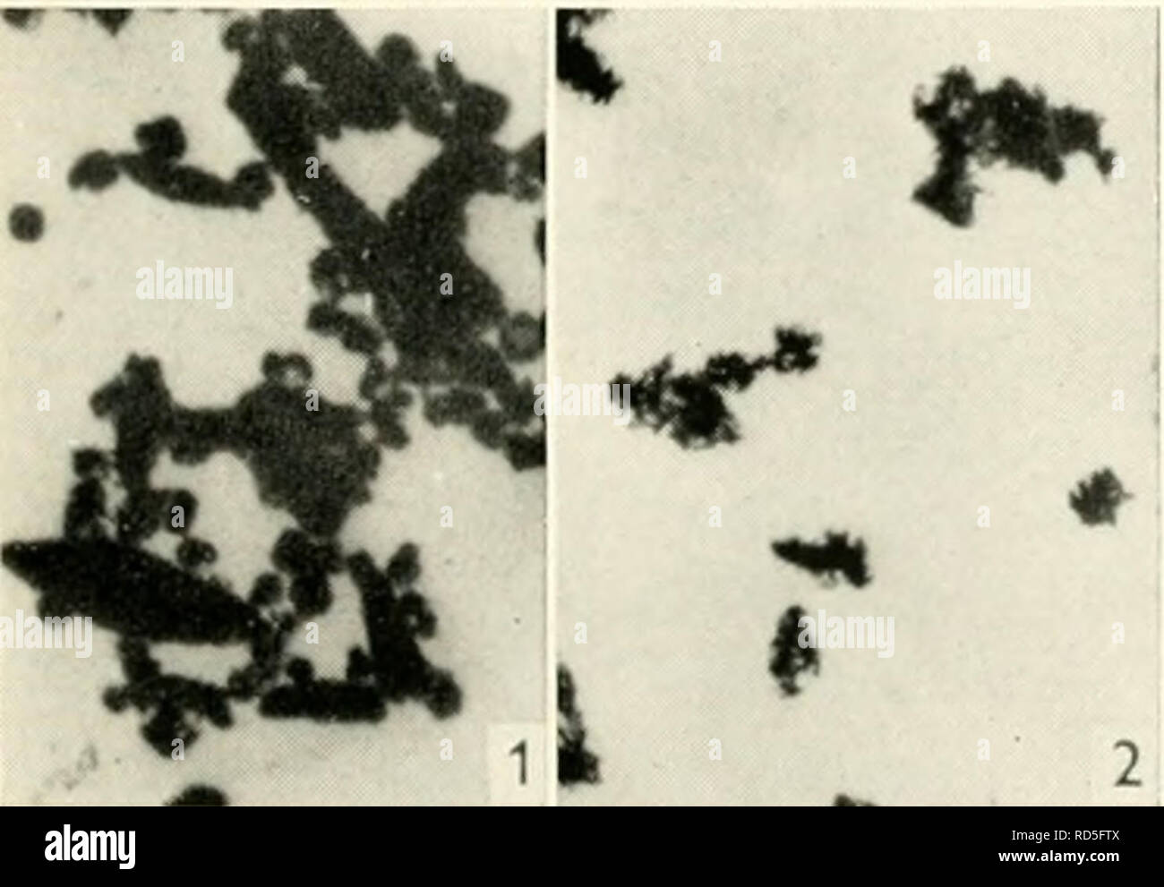
Gelatin silver process hi-res stock photography and images - Alamy

How it looks Jelly Gelatin at Amazing x500 x1500 Zoom 4K - Under the  Microscope Series - YouTube
How it looks Jelly Gelatin at Amazing x500 x1500 Zoom 4K - Under the Microscope Series - YouTube

Characterization of gelatin nanofiber prepared from gelatin–formic acid  solution - ScienceDirect
Characterization of gelatin nanofiber prepared from gelatin–formic acid solution - ScienceDirect

Light microscopic pictures of gelatin microspheres with diameters of... |  Download Scientific Diagram
Light microscopic pictures of gelatin microspheres with diameters of... | Download Scientific Diagram
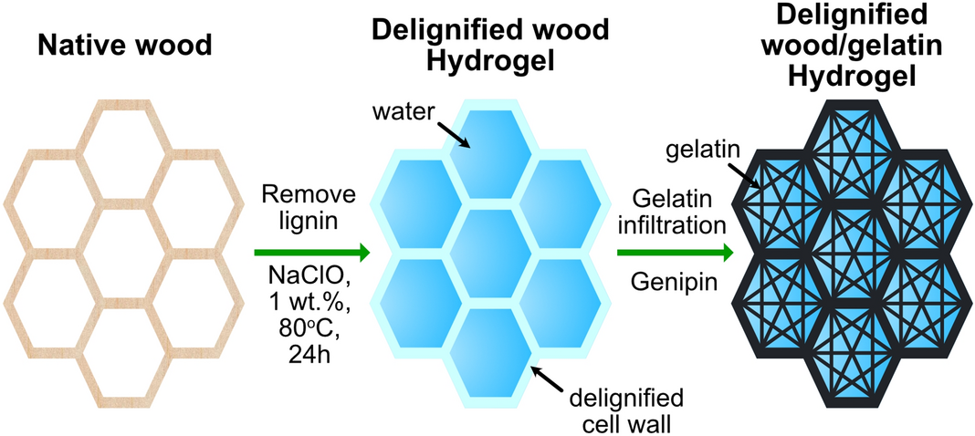
High strength and low swelling composite hydrogels from gelatin and  delignified wood | Scientific Reports
High strength and low swelling composite hydrogels from gelatin and delignified wood | Scientific Reports

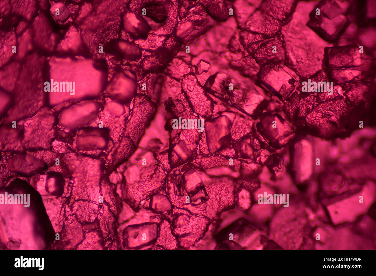
The Microscopic World. Fruit Jelly Under The Microscope. Stock Photo,  Picture and Royalty Free Image. Image 70207457.
The Microscopic World. Fruit Jelly Under The Microscope. Stock Photo, Picture and Royalty Free Image. Image 70207457.

Mannosylated gelatin microspheres as visualized by scanning electron... |  Download Scientific Diagram
Mannosylated gelatin microspheres as visualized by scanning electron... | Download Scientific Diagram
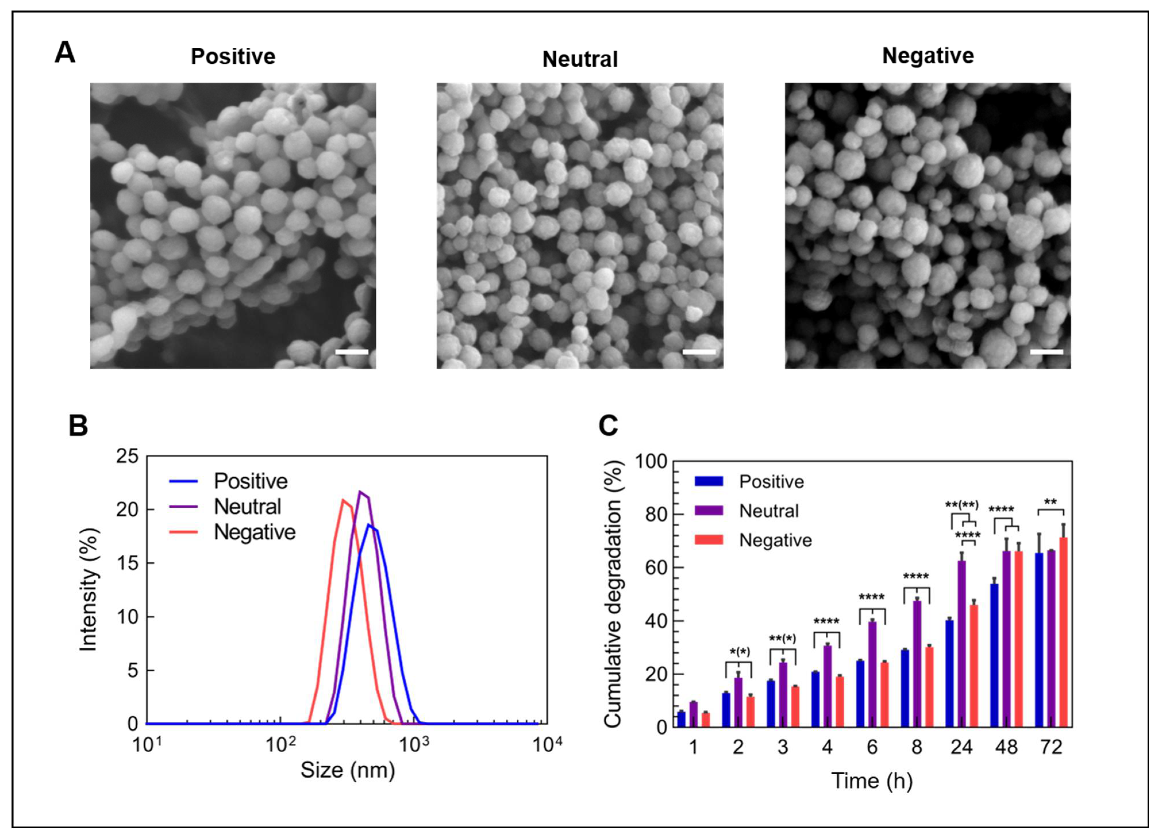
Nanomaterials | Free Full-Text | Gelatin Nanoparticles for Complexation and  Enhanced Cellular Delivery of mRNA
Nanomaterials | Free Full-Text | Gelatin Nanoparticles for Complexation and Enhanced Cellular Delivery of mRNA

Cryo-scanning electron microscopy (×5,000) of gelatin gels from poultry...  | Download Scientific Diagram
Cryo-scanning electron microscopy (×5,000) of gelatin gels from poultry... | Download Scientific Diagram
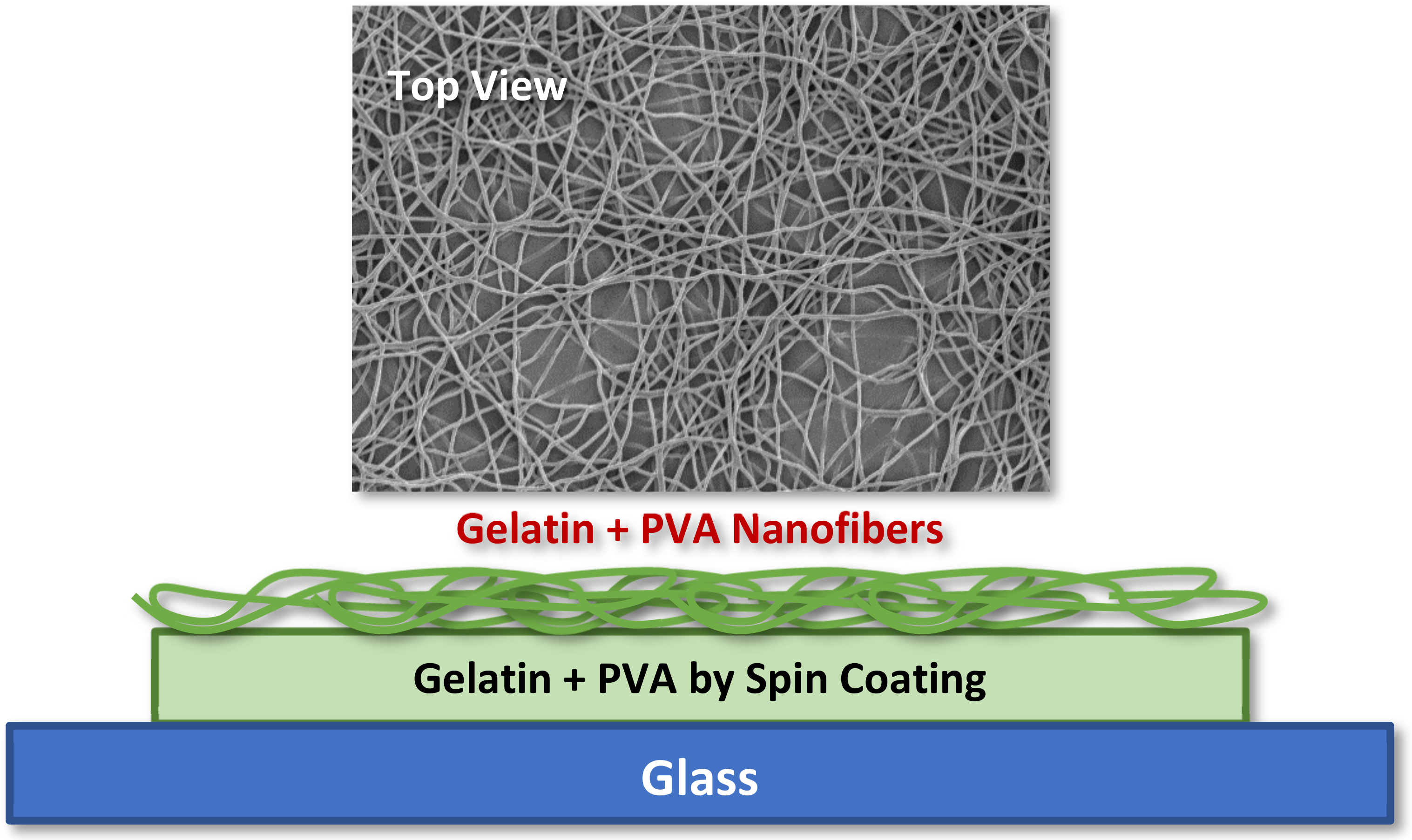
Polymers | Free Full-Text | Fabrication of Gelatin Nanofibers by  Electrospinning—Mixture of Gelatin and Polyvinyl Alcohol
Polymers | Free Full-Text | Fabrication of Gelatin Nanofibers by Electrospinning—Mixture of Gelatin and Polyvinyl Alcohol

Scanning electron microscopy images of gelatin fi lm. a, Pure gelatin... |  Download Scientific Diagram
Scanning electron microscopy images of gelatin fi lm. a, Pure gelatin... | Download Scientific Diagram

Scanning electron microscope of the surface of gelatin and collagen... |  Download Scientific Diagram
Scanning electron microscope of the surface of gelatin and collagen... | Download Scientific Diagram

Scanning electron microscope images of scaffolds made of Gelatin 5% w/v...  | Download Scientific Diagram
Scanning electron microscope images of scaffolds made of Gelatin 5% w/v... | Download Scientific Diagram

Scanning electron microscope (SEM) images of gelatine encapsulated C.... |  Download Scientific Diagram
Scanning electron microscope (SEM) images of gelatine encapsulated C.... | Download Scientific Diagram

The Microscopic World. Fruit jelly under the microscope Stock Photo - Alamy

Camera image of gelatine structure and optical microscope images of the...  | Download Scientific Diagram
Camera image of gelatine structure and optical microscope images of the... | Download Scientific Diagram

Optical microscope images of gelatin microspheres (GMs) in (A) dry and... |  Download Scientific Diagram
Optical microscope images of gelatin microspheres (GMs) in (A) dry and... | Download Scientific Diagram

Figure 7 from Microencapsulation of oil droplets using cold water fish  gelatine/gum arabic complex coacervation by membrane emulsification |  Semantic Scholar
Figure 7 from Microencapsulation of oil droplets using cold water fish gelatine/gum arabic complex coacervation by membrane emulsification | Semantic Scholar
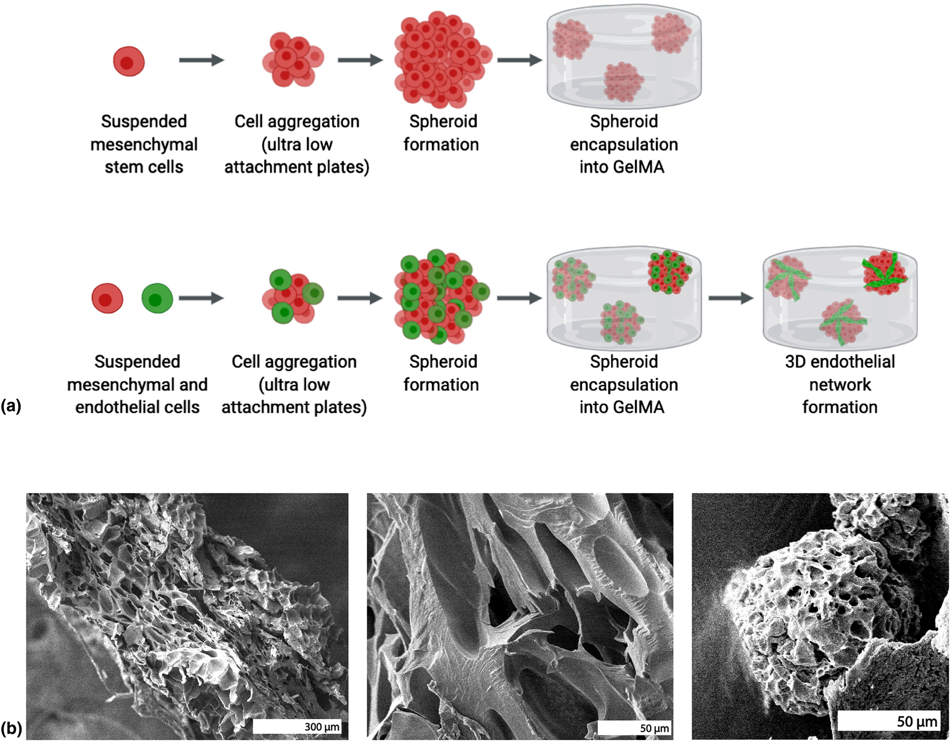
Photo-crosslinked gelatin methacrylate hydrogels with mesenchymal stem cell  and endothelial cell spheroids as soft tissue substitutes | Journal of  Materials Research | Cambridge Core
Photo-crosslinked gelatin methacrylate hydrogels with mesenchymal stem cell and endothelial cell spheroids as soft tissue substitutes | Journal of Materials Research | Cambridge Core
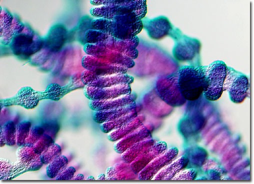
Molecular Expressions Microscopy Primer: Specialized Microscopy Techniques  - Differential Interference Contrast Image Gallery - Aurelia Jellyfish  Sensory Organs (Tentaculocysts)
Molecular Expressions Microscopy Primer: Specialized Microscopy Techniques - Differential Interference Contrast Image Gallery - Aurelia Jellyfish Sensory Organs (Tentaculocysts)

Morphology and microstructure of gelatin microspheres were observed via...  | Download Scientific Diagram
Morphology and microstructure of gelatin microspheres were observed via... | Download Scientific Diagram