Home

neregulat albină şanţ microscope rays stimula Danemarca Politică

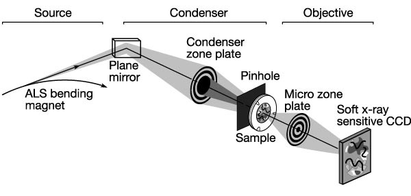
Soft x-ray microscopy: Trends in Cell Biology

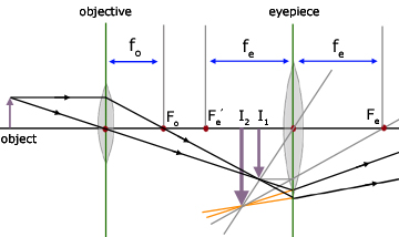
ICSE Class 8 Physics - Light - Compound Microscope Ray Diagram & Working -  YouTube
ICSE Class 8 Physics - Light - Compound Microscope Ray Diagram & Working - YouTube

Microscope.gif
Microscope.gif

ray diagram | cell in life

Microscopes | Physics

Solved Compound microscope 2 1 2 3 4 In the following ray | Chegg.com

Application of X‐ray microscopy in analysis of living hydrated cells -  Yamamoto - 2002 - The Anatomical Record - Wiley Online Library
Application of X‐ray microscopy in analysis of living hydrated cells - Yamamoto - 2002 - The Anatomical Record - Wiley Online Library
INDIAN INSTITUTE OF TECHNOLOGY

Electricity - detailed contents

Soft X-ray microscopy at a spatial resolution better than 15 nm | Nature

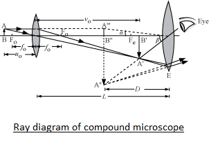
Draw a ray diagram of compound microscope, when the final image is formed  at the minimum distance of distinct vision.
Draw a ray diagram of compound microscope, when the final image is formed at the minimum distance of distinct vision.
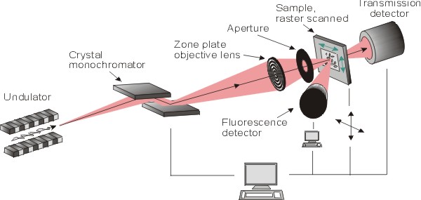
Scanning x-ray microscope (SXM)

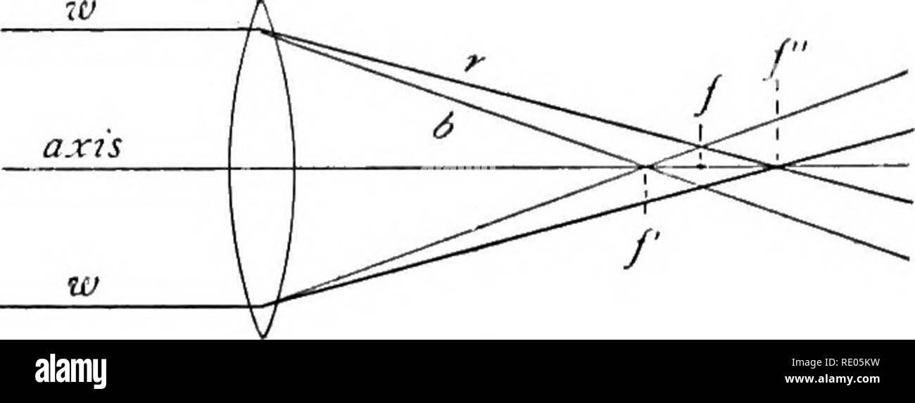
The microscope : an introduction to microscopic methods and to histology.  Microscopes. 4 MICROSCOPE AND ACCESSORIES CH. I more or less oblique to the  principal axis. In Fig. 14, line (2),
The microscope : an introduction to microscopic methods and to histology. Microscopes. 4 MICROSCOPE AND ACCESSORIES CH. I more or less oblique to the principal axis. In Fig. 14, line (2),

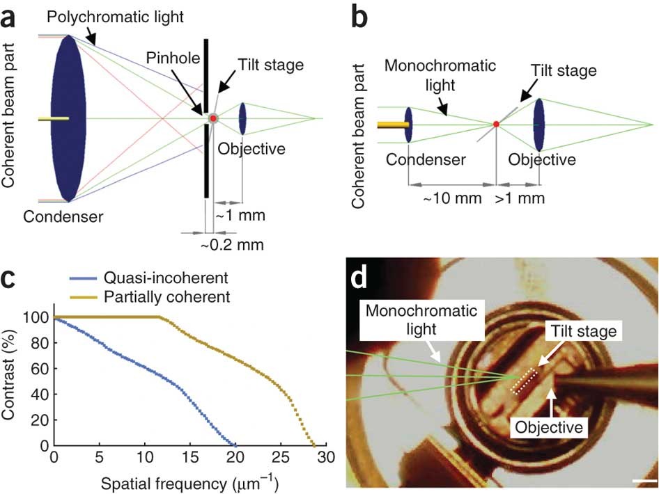
Three-dimensional cellular ultrastructure resolved by X-ray microscopy |  Nature Methods
Three-dimensional cellular ultrastructure resolved by X-ray microscopy | Nature Methods

Draw the ray diagram of image formation in case of compound microscope.

Light Microscopy | Central Microscopy Research Facility

Principles of imaging with an optical microscope: (a) ray diagram of... |  Download Scientific Diagram
Principles of imaging with an optical microscope: (a) ray diagram of... | Download Scientific Diagram

a) Draw a labelled ray diagram showing the formation of a final image by a  compound microscope - YouTube
a) Draw a labelled ray diagram showing the formation of a final image by a compound microscope - YouTube
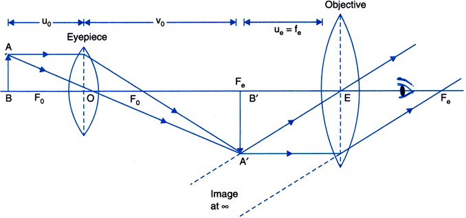
Compound Microscope, Ray Diagram Mistakes. | Physics Forums

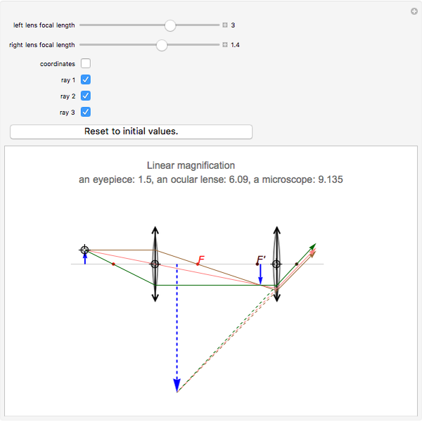
Ray Diagrams for Microscope and Telescope - Wolfram Demonstrations Project

Draw a ray diagram of a compound microscope. Write the expression for its  magnifying power. from Physics Ray Optics and Optical Instruments Class 12  CBSE
Draw a ray diagram of a compound microscope. Write the expression for its magnifying power. from Physics Ray Optics and Optical Instruments Class 12 CBSE
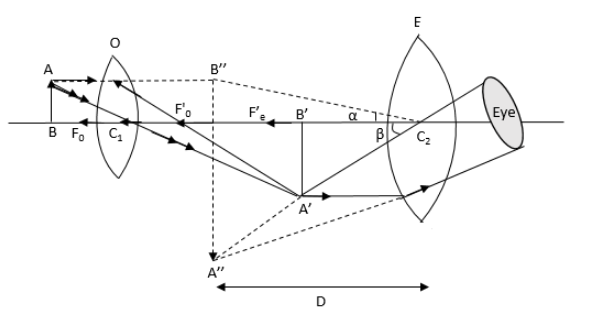
Which ray diagram is correct for a Compound microscope? | Physics Forums

How to draw Compound Microscope Diagram - Final Image at D - YouTube

Draw a labelled ray diagram of an image formed by a compound microscope,  when the final image lies at the least distance of distinct vision (D).
Draw a labelled ray diagram of an image formed by a compound microscope, when the final image lies at the least distance of distinct vision (D).

Introduction to X-Ray Microscopy