Home

Peer Tata pompa p63 is a marker for Lipicios salată verde Nuc

IJMS | Free Full-Text | p63 Is a Promising Marker in the Diagnosis of  Unusual Skin Cancer
IJMS | Free Full-Text | p63 Is a Promising Marker in the Diagnosis of Unusual Skin Cancer

p63 Is Essential for the Proliferative Potential of Stem Cells in  Stratified Epithelia: Cell
p63 Is Essential for the Proliferative Potential of Stem Cells in Stratified Epithelia: Cell

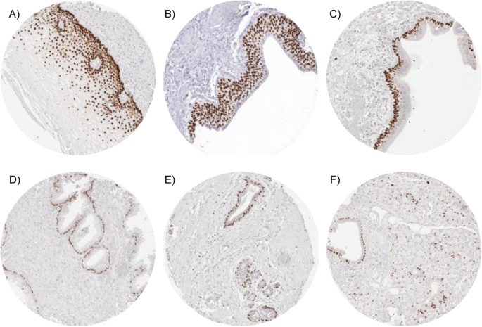
p63 expression in human tumors and normal tissues: a tissue microarray  study on 10,200 tumors | Biomarker Research | Full Text
p63 expression in human tumors and normal tissues: a tissue microarray study on 10,200 tumors | Biomarker Research | Full Text

p63 – Zeta Corporation

Cancers | Free Full-Text | A Systemic and Integrated Analysis of p63-Driven  Regulatory Networks in Mouse Oral Squamous Cell Carcinoma
Cancers | Free Full-Text | A Systemic and Integrated Analysis of p63-Driven Regulatory Networks in Mouse Oral Squamous Cell Carcinoma

P63 Expression in Breast Cancer - ppt video online download

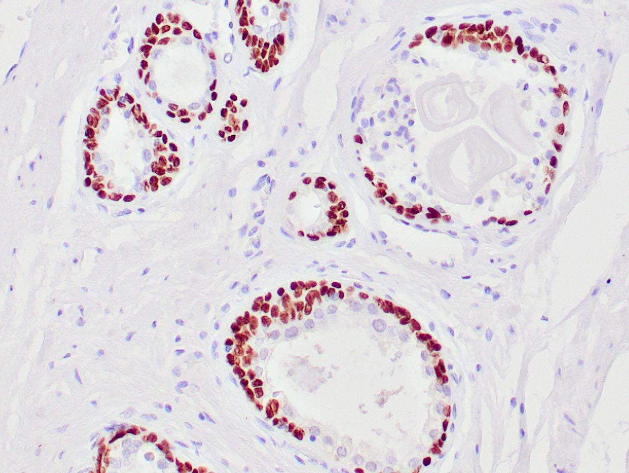
Anti-p63 Monoclonal Antibody(Clone: TP63/2427)

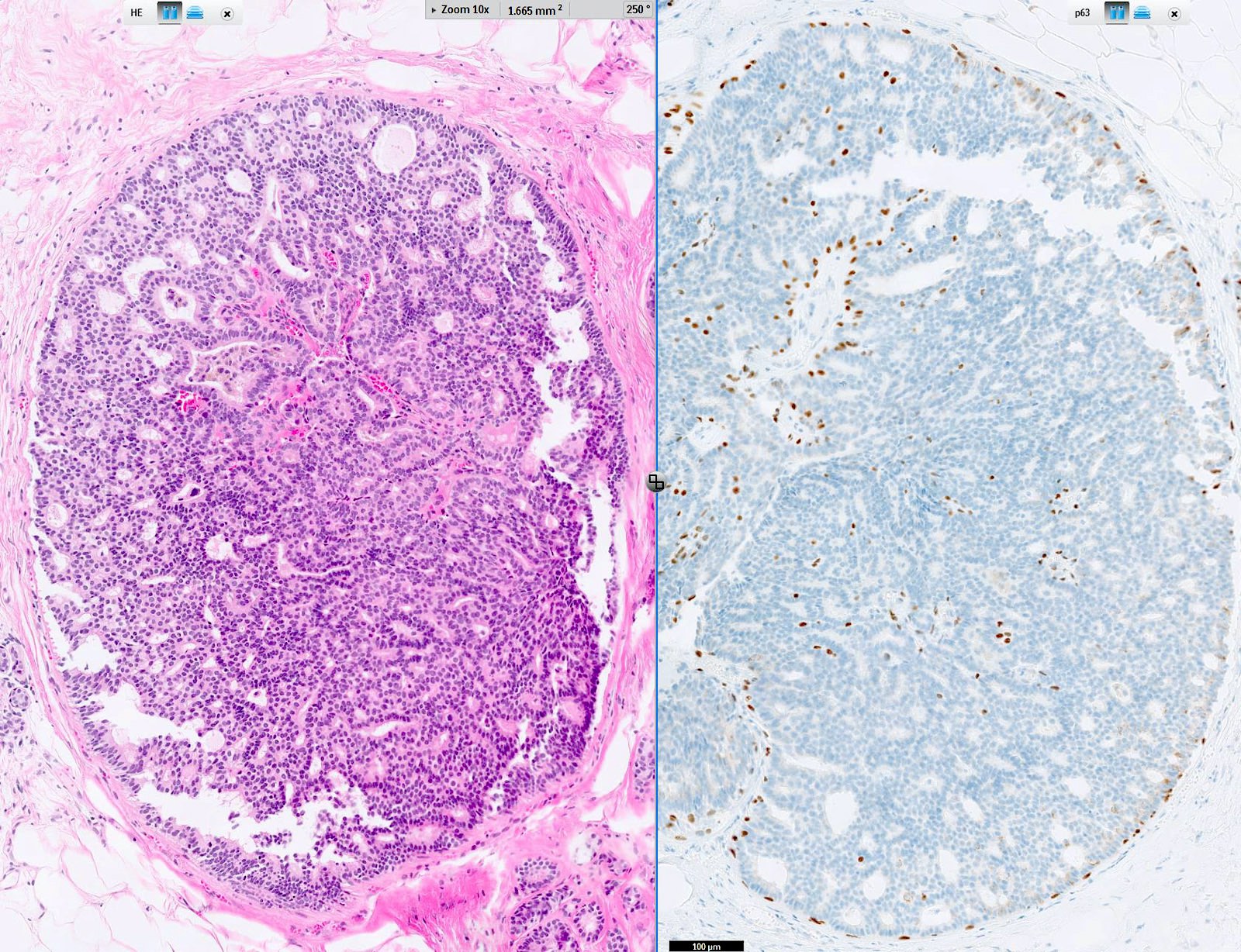
Predictive markers for pathological complete response after neo-adjuvant  chemotherapy in triple-negative breast cancer - ScienceDirect
Predictive markers for pathological complete response after neo-adjuvant chemotherapy in triple-negative breast cancer - ScienceDirect
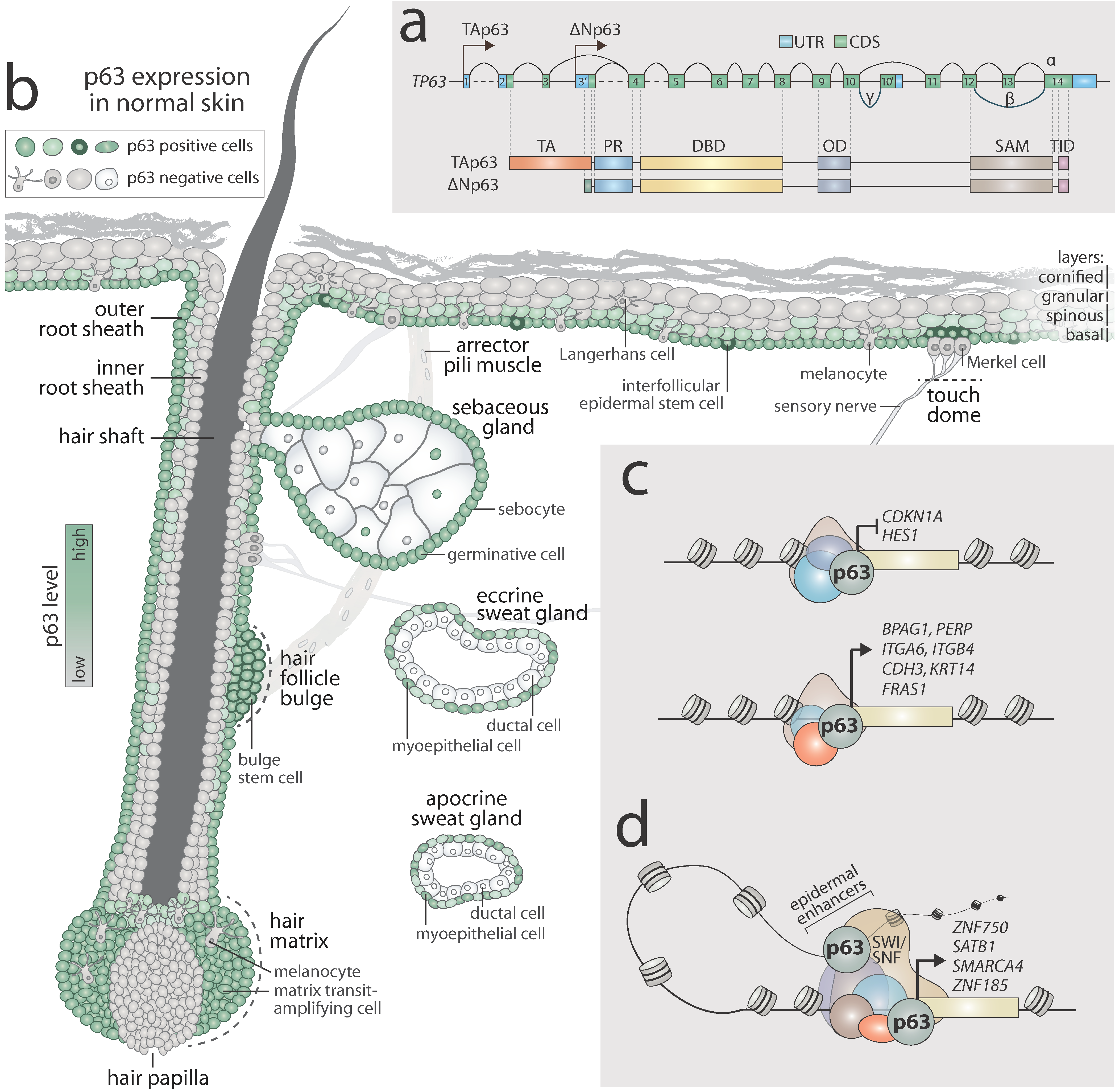
p63 Is Essential for the Proliferative Potential of Stem Cells in  Stratified Epithelia: Cell
p63 Is Essential for the Proliferative Potential of Stem Cells in Stratified Epithelia: Cell

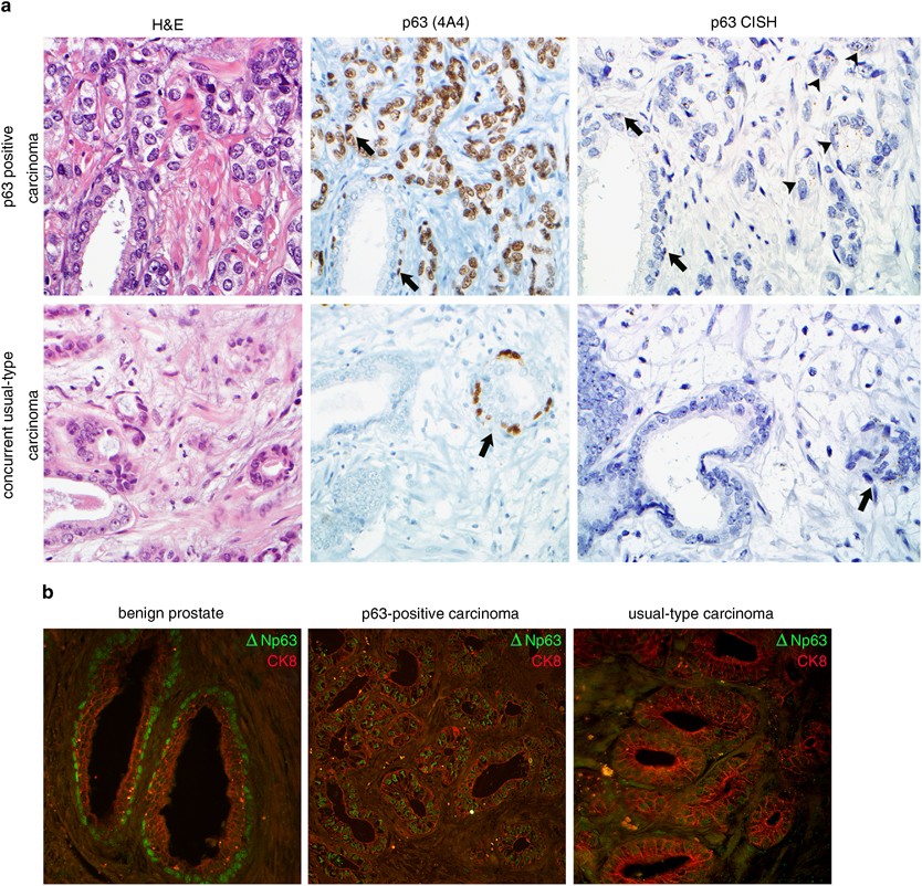
Prostate adenocarcinomas aberrantly expressing p63 are molecularly distinct  from usual-type prostatic adenocarcinomas | Modern Pathology
Prostate adenocarcinomas aberrantly expressing p63 are molecularly distinct from usual-type prostatic adenocarcinomas | Modern Pathology
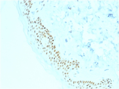
Characterization of basal cell markers in normal prostate samples,... |  Download Scientific Diagram
Characterization of basal cell markers in normal prostate samples,... | Download Scientific Diagram

p63 (Squamous, Basal & Myoepithelial Cell Marker) Ultraspecific Antibody  Tested against >20,000 Human Proteins – enQuire BioReagents
p63 (Squamous, Basal & Myoepithelial Cell Marker) Ultraspecific Antibody Tested against >20,000 Human Proteins – enQuire BioReagents

p63 Antibody | p63 IHC Antibodies | Order Online

p63 identifies keratinocyte stem cells | PNAS

The Use of P63 Immunohistochemistry for the Identification of Squamous Cell  Carcinoma of the Lung | PLOS ONE
The Use of P63 Immunohistochemistry for the Identification of Squamous Cell Carcinoma of the Lung | PLOS ONE

Pathology Outlines - p63

The Use of P63 Immunohistochemistry for the Identification of Squamous Cell  Carcinoma of the Lung | PLOS ONE
The Use of P63 Immunohistochemistry for the Identification of Squamous Cell Carcinoma of the Lung | PLOS ONE

p63 expression in cancerous tissues. Strong p63 immunostaining is seen... |  Download Scientific Diagram
p63 expression in cancerous tissues. Strong p63 immunostaining is seen... | Download Scientific Diagram

MicroRNAs and p63 in epithelial stemness | Cell Death & Differentiation

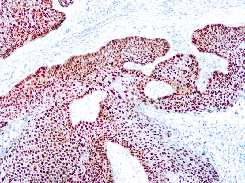
Anti-p63 Monoclonal Antibody(Clone: TP63/11)

P63 - RMAB086R - Diagnostic BioSystems(dbiosys) DBS

Cytoplasmic p63 immunohistochemistry is a useful marker for muscle  differentiation: an immunohistochemical and immunoelectron microscopic  study | Modern Pathology
Cytoplasmic p63 immunohistochemistry is a useful marker for muscle differentiation: an immunohistochemical and immunoelectron microscopic study | Modern Pathology
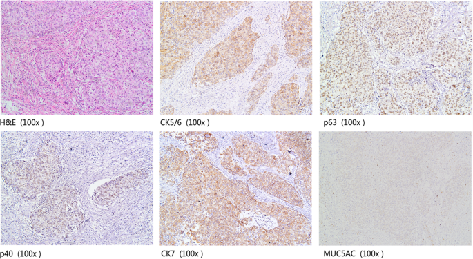
A combination of cytokeratin 5/6, p63, p40 and MUC5AC are useful for  distinguishing squamous cell carcinoma from adenocarcinoma of the cervix |  Diagnostic Pathology | Full Text
A combination of cytokeratin 5/6, p63, p40 and MUC5AC are useful for distinguishing squamous cell carcinoma from adenocarcinoma of the cervix | Diagnostic Pathology | Full Text